You may know of dental implants as one of the most definitive replacement options for missing tooth structures. However, not all individuals are ideal candidates for receiving conventional dental implants. In situations where the existing bone structure is insufficient, alternative treatment options might be necessary. One such alternative treatment option is rehabilitation with pterygoid implants, which are long implants placed in the bones surrounding the upper jaw.
Before you proceed, do go through the list of frequently encountered dental terminologies presented below to help you through this discussion.
GLOSSARY OF FREQUENTLY USED DENTAL TERMINOLOGIES:
DENTAL TERM
MEANING
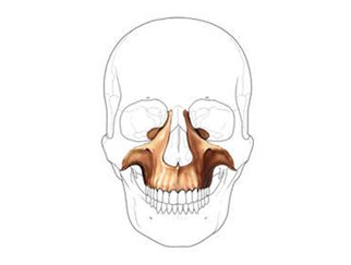
Maxilla
The jaw or jawbone, specifically the upper jaw in most vertebrates. In humans it also forms part of the nose and eye socket.

Maxillary tuberosity
A rounded eminence that marks the rearmost extent of maxilla.

Palatine bone
The bone situated at the back of the nasal cavity between the maxilla and the pterygoid process of the sphenoid bone.
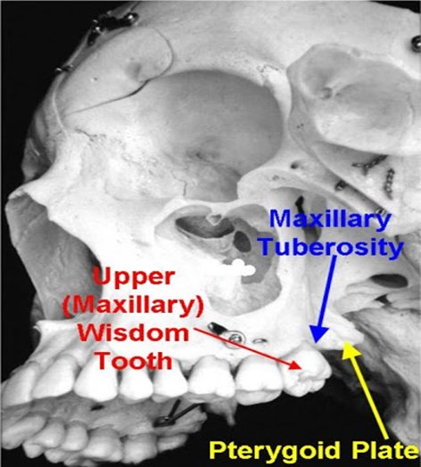
Pterygoid bone
The pterygoid is a paired bone located behind the palatine bones.
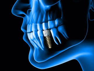
Dental Implant
The dental implant is a surgical component, shaped like the root of a natural tooth, that is placed within the bone of the maxilla or mandible to support an artificial tooth like restoration.

Abutment
The dental implant abutment is that part of the fixture that connects the artificial crown to the underlying dental implant.
Anterior
Front or up ahead
Posterior
Behind
Prosthesis
An artificial replacement of a missing natural body part, which in our case is the tooth.
Provisional Restoration
The artificial restoration that is placed temporarily till healing completes or the final restoration is ready is called the provisional or temporary restoration.
Cantilever
Technically, a cantilever means that a long beam is supported only on one end and is free on the other end.
Atrophic bone
Severe reduction in bone structure, commonly seen in older individuals.
Immediate Loading
The process of placement of a prosthesis immediately after the implant surgery to assist in esthetics, function, rehabilitation and recovery.
Denture Conversion
The process of using a premade denture for immediate loading after the completion of surgery.
WHAT ARE PTERYGOID IMPLANTS?
Pterygoid implants were first described by Tulsane in 1989 as a solution for atrophic posterior maxilla. These are long implants (15-20mm) that pass through the maxillary tuberosity and palatine bone and then engage the pterygoid process of sphenoid bone. The implant enters in the maxillary first or second molar region and follows an oblique direction proceeding posteriorly to find anchorage in the pterygoid fossa of the sphenoid bone.

WHEN ARE PTERYGOID IMPLANTS INDICATED?
- In cases with limited bone quantity (atrophic maxilla)
- When the maxillary sinus lining is close to the alveolar bone
- As an alternative to bone augmentation
BENEFITS OF ZYGOMATIC IMPLANTS:
- Less invasive:
The biggest advantage with pterygoid implant is that is avoids complex procedures like bone augmentation and sinus lifts. Bone grafts are often quite unpredictable and are associated with donor site morbidity, which can be prevented with the use of pterygoid implants. - horter treatment time:
The pterygoid implant placement and prosthesis delivery can be done on the same day unlike grafting procedures, thus shortening the treatment time to a large extent. - Good success rates:
The success rate for pterygoid implants has been reported from 71% to nearly 100%. In a study conducted by Bahat et al, the mean survival rate was noted to be 93%. - Better Chewing efficiency:
Since the implants are placed in posterior maxilla, greater extensions can be provided in the prosthesis without the need for distal cantilevers.
STEPS IN THE PROCEDURE:
The phases in the procedure can be simply explained with the following flow chart:
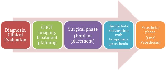
The planning for pterygoid implants is always best done in reverse. A properly executed treatment plan with CBCT images will help the clinician in deciding the optimum location for placing the implant. A through clinical evaluation of the patient’s oral conditions and systemic health is the first step, followed by virtual planning and obtaining measurements of the existing bone structures.
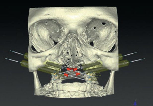
Potential implant sites are determined with CBCT images (as shown in figure)
The surgical phase involves the following steps:
SURGICAL PHASE:
1. Selection of optimum entry point in the alveolar crest based on the scanned images (Usually 3-4mm anterior to the maxillary tuberosity)
2. Flap reflection and placement of implants.
3. Verification of implant positions with a radiograph or volumetric image
4. Flap closure.
The surgical procedure can also be carried out with the use of 3D printed guided stents which assist in precise placement of implants according to the plan.
The prosthetic phase begins soon after surgery where a provisional restoration is fabricated. In completely edentulous patients, a previously made denture is converted intra-orally into temporary implant prosthesis. This temporary denture will remain in place until the permanent prosthesis is fabricated.
PROSTHETIC PHASE (INTERMEDIATE AND FINAL):
1. Open tray impression with splinted impression copings
2. Jig verification of the implant positions
3. Jaw relation verification
4. Bridge design using CAD-CAM technology
5. Fabrication of a zirconia bridge/DMLS metal-ceramic bridge (in partially edentulous)/ titanium framework
6. Trial and finishing
7. Final fixation in the mouth
Conclusion:
Pterygoid dental implants can be considered as a suitable treatment option for replacement of teeth in the posterior maxilla. They offer several advantages over bone grafts/sinus lifts and demonstrate remarkable success rates.


